植物生态学报 ›› 2008, Vol. 32 ›› Issue (3): 690-697.DOI: 10.3773/j.issn.1005-264x.2008.03.019
李荣峰1,2( ), 蔡妙珍1, 刘鹏1,*(), 徐根娣1, 陈敏燕1, 梁和2
收稿日期:2006-12-04
接受日期:2007-07-11
出版日期:2008-12-04
发布日期:2008-05-30
通讯作者:
刘鹏
作者简介:*E-mail:sky79@zjnu.cn基金资助:
LI Rong-Feng1,2(), CAI Miao-Zhen1, LIU Peng1,*(), XU Gen-Di1, CHEN Min-Yan1, LIANG He2
Received:2006-12-04
Accepted:2007-07-11
Online:2008-12-04
Published:2008-05-30
Contact:
LIU Peng
摘要:
设置不同的Al3+浓度(0、25、50、100、200、400 μmol·L -1)和培养时间(12、24 h),研究了边缘细胞活性和大豆(Glycine max)根中过氧化氢酶(CAT)、过氧化物酶(POD)、超氧化物歧化酶(SOD)随Al3+浓度及处理时间变化的规律,并通过Hoechst33342-PI双重荧光染色、梯状DNA(即DNA ladder)分析和末端脱氧核糖核酸转移酶介导的dUTP切口末端标记(即TUNEL原位标记)检测,研究了Al3+对大豆根边缘细胞程序性死亡诱导的生理生态作用。结果表明,Al3+胁迫能诱导边缘细胞的死亡,随着Al3+浓度的升高和处理时间的延长,细胞死亡率增加。通过Hoechst33342-PI双重荧光染色、DNA ladder分析和TUNEL原位标记,检测到Al3+胁迫下发生程序性死亡的边缘细胞。其表现为:在400 μmol·L -1 Al3+诱导大豆根24 h时, 核酸电泳显示细胞DNA发生特异性降解并形成阶梯状电泳条带(DNA ladder),用TUNEL原位标记检测200和400 μmol·L -1 Al3+ 处理12 h后的大豆根边缘细胞,发现DNA的3'-OH端被原位特异标记,二氨基联苯胺(DAB)显色后,细胞核为阳性或强阳性。同时,高浓度Al3+(>100 μmol·L-1)处理下,CAT、POD和SOD活性均有不同程度的下降,CAT和SOD的活性也随处理时间的延长而降低。说明在Al3+胁迫下边缘细胞的死亡可能是一种程序性死亡形式,高浓度Al3+胁迫下,通过诱导活性氧在细胞体内的产生和累积而导致细胞凋亡,此过程是其对逆境胁迫所作出的生理生态防御性应答方式之一。
李荣峰, 蔡妙珍, 刘鹏, 徐根娣, 陈敏燕, 梁和. Al3+对大豆根边缘细胞程序性死亡诱导的生理生态作用. 植物生态学报, 2008, 32(3): 690-697. DOI: 10.3773/j.issn.1005-264x.2008.03.019
LI Rong-Feng, CAI Miao-Zhen, LIU Peng, XU Gen-Di, CHEN Min-Yan, LIANG He. PHYTOECOLOGICAL EFFECT OF Al3+ ON THE INDUCTIVITY OF PROGRAMMED CELL DEATH OF BORDER CELLS IN SOYBEAN ROOT. Chinese Journal of Plant Ecology, 2008, 32(3): 690-697. DOI: 10.3773/j.issn.1005-264x.2008.03.019
| 处理时间 Treatment time (h) | Al3+ 浓度 Al3+ concentration (μmol·L-1) | ||||||||
|---|---|---|---|---|---|---|---|---|---|
| 0 | 50 | 100 | 200 | 400 | |||||
| 12 | 84.39±3.68ab | 79.64±6.52abc | 72.68±7.36bc | 71.26±6.41c | 67.49±15.30cd | ||||
| 24 | 85.78±2.72a | 72.00±5.38bc | 66.87±0.29cd | 57.33±4.08de | 49.54±5.79e | ||||
表1 大豆边缘细胞存活率随Al3+浓度和处理时间的变化(平均值±标准误差)
Table 1 Viabilities (%) of soybean root border cells treated with different Al3+ concentrations and treatment time (Mean±SE)
| 处理时间 Treatment time (h) | Al3+ 浓度 Al3+ concentration (μmol·L-1) | ||||||||
|---|---|---|---|---|---|---|---|---|---|
| 0 | 50 | 100 | 200 | 400 | |||||
| 12 | 84.39±3.68ab | 79.64±6.52abc | 72.68±7.36bc | 71.26±6.41c | 67.49±15.30cd | ||||
| 24 | 85.78±2.72a | 72.00±5.38bc | 66.87±0.29cd | 57.33±4.08de | 49.54±5.79e | ||||
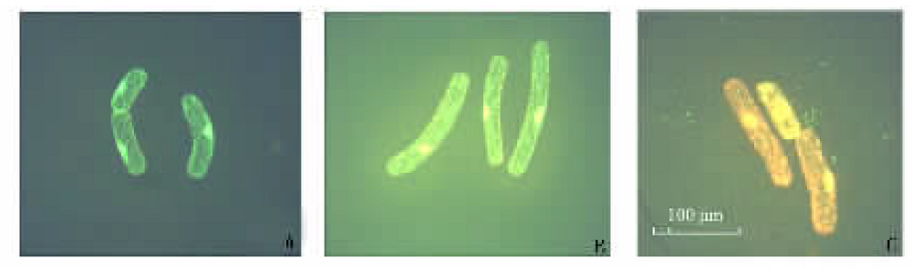
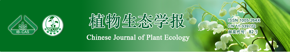
图1 大豆边缘细胞Hoechst33342-PI双重染色荧光效果 A:活的边缘细胞 Live root border cells B:程序性死亡的大豆边缘细胞 Soybean root border cells of programmed cell death C:坏死细胞或晚期的凋亡细胞 Dead cells or later-phase apoptosis cells in soybean root border cells
Fig.1 Hoechst33342-PI double staining results in soybean root border cells
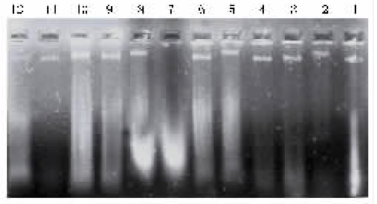
图2 Al3+处理12和24 h后大豆边缘细胞DNA的凝胶电泳图谱 1~6:分别表示0、25、50、100、200、400 μmol·L -1 Al3+处理12 h Treated with 0, 25, 50, 100, 200 and 400 μmol·L -1 Al3+ for 12 h, respectively 7~12:分别表示0、25、50、100、200、400 μmol·L -1 Al3+处理24 h Treated with 0, 25, 50, 100, 200 and 400 μmol·L -1 Al3+ for 24 h, respectively
Fig.2 DNA band patterns are visualized in soybean root border cells treated with different Al3+ concentrations for 12 h and 24 h, respectively
图3 Al3+诱导大豆边缘细胞程序性死亡的TUNEL检测结果 A、B、C、D、E、F:分别表示0、25、50、100、200和400 μmol·L -1 Al3+处理12 h后的边缘细胞 Showing the root border cells treated with 0, 25, 50, 100, 200 and 400 μmol·L -1 Al3+ for 12 h, respectively
Fig.3 In situ detection with the TUNEL reaction for soybean root border cells exposed to Al3+
| 处理时间 Treatment time (h) | Al3+ 浓度 Al3+ concentration (μmol·L-1) | CAT活性 CAT activity (U·mg-1 Pr) | POD活性 POD activity (Δ470·mg-1Pr·min-1) | SOD 活性 SOD activity (U·mg-1Pr)(×103) |
|---|---|---|---|---|
| 12 | 0 | 265.9±76.52a | 144.5±87.65a | 27.7±2.68ab |
| 25 | 291.1±41.92a | 148.1±32.58a | 30.5±11.04ab | |
| 50 | 283.3±4.45a | 151.9±12.84a | 31.2±4.09ab | |
| 100 | 272.0±113.6a | 139.5±34.01a | 36.2±13.70a | |
| 200 | 255.2±30.23a | 118.2±8.03a | 30.7±1.35ab | |
| 400 | 159.0±43.93b | 114.5±12.59a | 28.0±4.90ab | |
| 24 | 0 | 67.68±18.65d | 152.8±7.92a | 20.5±9.87bc |
| 25 | 106.2±13.08bcd | 158.2±30.98a | 21.4±0.51bc | |
| 50 | 147.7±45.46bc | 160.5±23.4a | 31.1±8.47ab | |
| 100 | 113.2±7.96bcd | 157.4±12.73a | 23.3±0.55abc | |
| 200 | 77.10±0.17cd | 163.5±2.15a | 21.4±3.45bc | |
| 400 | 138.5±21.28bcd | 151.9±8.25a | 14.3±3.08c |
表2 Al3+对大豆根尖CAT、POD和SOD酶活性的影响(平均值±标准误差)
Table 2 Effects of different Al3+ concentrations on the activities of CAT, POD and SOD in soybean root tips (Mean±SE)
| 处理时间 Treatment time (h) | Al3+ 浓度 Al3+ concentration (μmol·L-1) | CAT活性 CAT activity (U·mg-1 Pr) | POD活性 POD activity (Δ470·mg-1Pr·min-1) | SOD 活性 SOD activity (U·mg-1Pr)(×103) |
|---|---|---|---|---|
| 12 | 0 | 265.9±76.52a | 144.5±87.65a | 27.7±2.68ab |
| 25 | 291.1±41.92a | 148.1±32.58a | 30.5±11.04ab | |
| 50 | 283.3±4.45a | 151.9±12.84a | 31.2±4.09ab | |
| 100 | 272.0±113.6a | 139.5±34.01a | 36.2±13.70a | |
| 200 | 255.2±30.23a | 118.2±8.03a | 30.7±1.35ab | |
| 400 | 159.0±43.93b | 114.5±12.59a | 28.0±4.90ab | |
| 24 | 0 | 67.68±18.65d | 152.8±7.92a | 20.5±9.87bc |
| 25 | 106.2±13.08bcd | 158.2±30.98a | 21.4±0.51bc | |
| 50 | 147.7±45.46bc | 160.5±23.4a | 31.1±8.47ab | |
| 100 | 113.2±7.96bcd | 157.4±12.73a | 23.3±0.55abc | |
| 200 | 77.10±0.17cd | 163.5±2.15a | 21.4±3.45bc | |
| 400 | 138.5±21.28bcd | 151.9±8.25a | 14.3±3.08c |
| [1] | Amako K, Chen GX, Asade K (1994). Separate assays specific for ascorbate peroxidase and guaiacol peroxidase and for the chloroplastic and cytosolic isozymes of ascorbate peroxidase in plants. Plant & Cell Physiology, 35,497-504. |
| [2] | Boscolo PRS, Menossi M, Jorqe RA (2003). Aluminum-induced oxidative stress in maize. Phytochemistry, 62,181-189. |
| [3] | Cakmak I, Horst WJ (1991). Effect of aluminum on lipid peroxidation, superoxide dismutase, catalase, and peroxidase activities in root tips of soybean (Glycine max). Physiologia Plantarum, 834,463-468. |
| [4] | Cai MZ (蔡妙珍), Liu P (刘鹏), Xu GD (徐根娣), Liu HH (刘海华) (2006). Effect of Al3+ toxicity on root border cells in vitro of buckwheat. Journal of Jiangsu University (Natural Science Edition) (江苏大学学报(自然科学版)), 27,293-298. (in Chinese with English abstract) |
| [5] | Cai MZ (蔡妙珍), Liu P (刘鹏), Xu GD (徐根娣), Liu WX (刘文秀), Gong CF (龚春风) (2007). Response of root border cells to Al3+ toxicity in soybean . Scientia Agricultura Sinica (中国农业科学), 40,271-276. (in Chinese with English abstract) |
| [6] |
Campbell A, Prasad KN, Bondy SC (1999). Aluminum-induced oxidative events in cell lines: glioma are more responsive than neuroblastoma. Free Radical Biology & Medicine, 26,1166-1171.
URL PMID |
| [7] |
de Jong AJ, Hoeberichts FA, Yakimova ET, Maximova E, Woltering EJ (2000). Chemical-induced apoptotic cell death in tomato cells: involvement of caspase-like proteases. Planta, 211,656-662.
URL PMID |
| [8] | de Jong AJ, Elena TY, Veneta MK, Ernist JW (2002). A critical role for ethylene in hydrogen peroxide release during programmed cell death in tomato suspension cells. Planta, 214,537-545. |
| [9] | Du X (杜幸), Liu P (刘鹏), Xu GD (徐根娣), Cai MZ (蔡妙珍) (2006). Response of root border cell in red cowpea to aluminum stress. Plant Nutrition and Fertilizer Science (植物营养与肥料学报), 12,722-726. (in Chinese with English abstract) |
| [10] | Feng YM (冯英明), Yu M (喻敏), Wen HY (温海洋), Zhang YH (张英慧), Xiao HD (萧洪东), Wang HZ (王惠珍), He LL (何丽烂), Liang HD (梁火娣) (2005). Influence of Al on cell viability and mucilage of root border cells of pea (Pisum sativum). Ecological Environment (生态环境), 14,695-699. (in Chinese with English abstract) |
| [11] |
Greenberg JT (1996). Programmed cell death: a way of life for plants. Proceedings of the National Academy of Sciences of the United States of America, 93,12094-12097.
DOI URL PMID |
| [12] | Hawes MC, Brigham LA (1992). Impact of root border cells on microbial populations in the rhizosphere. Advances in Plant Pathology, 8,l19-148. |
| [13] | Hawes MC, Gunawardena U, Miyasaka SC, Zhao XW (2000). The role of root border cells in plant defense. Trends in Plant Science, 5,128-133. |
| [14] | Hoeberichts FA, Woltering EJ (2002). Multiple mediators of plant programmed cell death: interplay of conserved cell death mechanisms and plant-specific regulators. Bioessays, 25,47-57. |
| [15] | Jones AW (2001). Programmed cell death in development and defense. Plant Physiology, 125,94-97. |
| [16] | Kochian LV (1995). Cellular mechanisms of aluminum toxicity and resistance in plants. Annual Review of Plant Physiology and Plant Molecular Biology, 46,237-260. |
| [17] |
Levine A, Pennell RI, Alvarez ME, Palmer R, Lamb C (1996). Calcium-mediated apoptosis in a plant hypersensitive disease resistance response. Current Biology, 6,427-437.
URL PMID |
| [18] | Lu XD (鲁旭东), Xie ZB (谢志兵) (2003). Programmed cell death in plant growth and development. Journal of Xiaogan University (孝感学院学报), 23 (6),53-56. (in Chinese with English abstract) |
| [19] | Matsumoto H (2000). Cell biology of aluminum toxicity and tolerance in higher plants. International Review of Cytology, 200,41-46. |
| [20] | McCabe PF, Leaver CJ (2000). Programmed cell death in cell cultures. Plant Molecular Biology, 44,359-368. |
| [21] | Mittler R, Lam E (1997). Characterization of nuclease activities and DNA fragmentation induced upon hypersensitive response cell death and mechanical stress. Plant Molecular Biology, 34,209-221. |
| [22] |
Miyasaka SC, Hawes MC (2001). Possible role of root border cells in detection and avoidance of aluminum toxicity. Plant Physiology, 125,1978-1987.
URL PMID |
| [23] | Ning SB, Wang L, Song YC (1999). Programmed cell death in plants: a new emerging research field. Development & Reproductive Biology, 8,71-100. |
| [24] | Ormerod MG, Sun XM, Brown D, Snowden RT, Cohen GM (1993). Quantification of apoptosis and necrosis by flow cytometry. Acta Oncologica, 32,417-424. |
| [25] | Orzaez D, Granell A (1997). DNA fragmentation is regulated by ethylene during carpel senescence in Pisum sativum. The Plant Journal, 11,137-144. |
| [26] | Pan JW (潘建伟) (2002). Biological Characters and Mechanisms of Aluminum Toxicity in the Root Tips and Border Cells of Barley (大麦根尖和边缘细胞铝毒生物学特性及其机理研究). PhD dissertation, Zhejiang University, Hangzhou,98-103. (in Chinese) |
| [27] | Pennell RI, Lamb C (1997). Programmed cell death in plants. The Plant Cell, 9,1157-1168. |
| [28] |
Richards KD, Schott EJ, Sharma YK, Davis KR, Gardner RC (1998). Aluminum induces oxidative stress genes in Arabidopsis thaliana. Plant Physiology, 116,409-418.
DOI URL PMID |
| [29] | Rosa AV, Maria C, Daniela V, Salvatore P, Ersilia M, Laura DG (2004). Production of reactive oxygen species, alteration of cytosolic ascorbate peroxidase, and impairment of mitochondrial metabolism are early events in heat shock-induced programmed cell death in tobacco bright-yellow cells. Plant Physiology, 134,1100-1112. |
| [30] | Sun YL (孙英丽), Zhao Y (赵允), Liu CX (刘春香), Zhai ZH (翟中和) (1999). Cytochrome c can induce programmed cell death in plant cells. Acta Botanica Sinica (植物学报), 41,379-383. (in Chinese with English abstract) |
| [31] |
Tamas L, Budlkova S, Huttova J, Mistrik I, Simonovicovicova M, Siroka B (2005). Aluminum-induced cell death of barley—root border cells is correlated with peroxidase- and oxalate oxidase-mediated hydrogen peroxide production. Plant Cell Reports, 24,189-194.
DOI URL PMID |
| [32] | Wang CB (王淳本) (2003). Applied Biochemistry and Molecular Biology Technology (实用生物化学与分子生物学实验技术) 2nd edn. Hubei Science and Technology Press, Wuhan, 226. (in Chinese) |
| [33] | Wang AG (王爱国), Luo GH (罗广华), Shao CB (邵从本), Wu SJ (吴淑君), Guo JY (郭俊彦) (1983). A study on the superoxide dismutase of soybean seeds. Acta Botanica Sinica (植物学报), 9,77-84. (in Chinese with English abstract) |
| [34] |
Wang H, Li J, Bostock RM, Gilchrist DG (1996). Apoptosis: a function paradigm for programmed plant cell death induced by a host-selective phytotoxin and invoked during development. The Plant Cell, 8,375-391.
URL PMID |
| [35] | Xu GD (徐根娣), Liu P (刘鹏), Zhou ZH (周志华) (2004). A progress of studies on development and functions of border cells. Chinese Agricultural Science Bulletin (中国农学通报), 20(5),28-32. (in Chinese with English abstract) |
| [36] | Yamaguchi Y, Yamamoto, Matsumoto H (1999). Cell death process initiated by a combination of aluminum and iron in suspension-cultured tobacco ( Nicotiana tabacum) cells: apoptosis-like cell death mediated by calcium and proteinase . Soil Science & Plant Nutrition, 45,647-657. |
| [37] | Yu M (喻敏), Cui ZX (崔志新), Wen HX (温海祥), Xiao HD (萧洪东), Zhang YH (张英慧), He LL (何丽烂), Liang HD (梁火娣) (2004). Root border cells-A recently defined population of alive cells in rhizophere. Journal of Huazhong Agricultural University (华中农业大学学报), 23,275-280. (in Chinese with English abstract) |
| [38] | Yuan YJ (元英进), Ge ZQ (葛志强), Wang YD (王艳东), Ma ZY (马振毅), Hu ZD (胡宗定) (2001). Ce 4+ induces apoptosis suspension culture of Taxus cuspidata cells . Journal of the Chinese Rare Earth Society (中国稀土学报), 19,357-361. (in Chinese with English abstract) |
| [39] | Zhang ZL (张志良) (1990). The Experimental Guide for Plant Physiology (植物生理学实验指导). Higher Education Press, Beijing, 210-212. (in Chinese) |
| [40] | Zhao YG (赵云罡), Xu JX (徐建兴) (2001). Mitochondrion, AOS and apoptosis. Progress in Biochemistry and Biophysics (生物化学与生物物理进展), 28,168-171. (in Chinese with English abstract) |
| [41] | Zhou N (周楠), Chen WR (陈文荣), Liu P (刘鹏), Xu GD (徐根娣) (2006). Biological characteristic and the response to aluminum toxicity of cucumber border cells. Acta Horticulturae Sinica (园艺学报), 33,1117-1120. (in Chinese with English abstract) |
| [42] |
Zhu MY, Ahn S, Matsumoto H (2003). Inhibition of growth and development of root border cells in wheat by Al. Physiologia Plantarum, 117,359-367.
URL PMID |
| [1] | 邓蓓 王晓锋 廖君. 环境胁迫影响三峡库区消落带草本和木本植物生理生态特征的整合分析[J]. 植物生态学报, 2024, 48(5): 623-637. |
| [2] | 余玉蓉, 吴浩, 高娅菲, 赵媛博, 李小玲, 卜贵军, 薛丹, 刘正祥, 武海雯, 吴林. 模拟氮沉降对鄂西南湿地泥炭藓生理及形态特征的影响[J]. 植物生态学报, 2023, 47(11): 1493-1506. |
| [3] | 祁鲁玉, 陈浩楠, 库丽洪·赛热别力, 籍天宇, 孟高德, 秦慧颖, 王宁, 宋逸欣, 刘春雨, 杜宁, 郭卫华. 基于植物功能性状的暖温带5种灌木幼苗生长策略[J]. 植物生态学报, 2022, 46(11): 1388-1399. |
| [4] | 叶子飘, 段世华, 安婷, 康华靖. 最大电子传递速率的确定及其对电子流分配的影响[J]. 植物生态学报, 2018, 42(4): 498-507. |
| [5] | 许红梅, 李进, 张元明. 水分条件对人工培养齿肋赤藓光化学效率及生理特性的影响[J]. 植物生态学报, 2017, 41(8): 882-893. |
| [6] | 刘盟盟, 贾丽, 程路芸, 张洪芹, 臧晓琳, 宝音陶格涛, 张汝民, 高岩. 冷蒿酚酸及其抗氧化防御酶活性对机械损伤的响应[J]. 植物生态学报, 2017, 41(2): 219-230. |
| [7] | 王丹, 乔匀周, 董宝娣, 葛静, 杨萍果, 刘孟雨. 昼夜不对称性与对称性升温对大豆产量和水分利用的影响[J]. 植物生态学报, 2016, 40(8): 827-833. |
| [8] | 尹本丰, 张元明. 冻融过程对荒漠区不同微生境下齿肋赤藓渗透调节物含量和抗氧化酶活力的影响[J]. 植物生态学报, 2015, 39(5): 517-529. |
| [9] | 龚容, 高琼. 叶片结构的水力学特性对植物生理功能影响的研究进展[J]. 植物生态学报, 2015, 39(3): 300-308. |
| [10] | 吴启美, 周启星. 大金发藓对土壤多氯联苯污染的生理生态响应[J]. 植物生态学报, 2015, 39(3): 275-282. |
| [11] | 彭东海,杨建波,李健,邢永秀,覃刘东,杨丽涛,李杨瑞. 间作大豆对甘蔗根际土壤细菌及固氮菌多样性的影响[J]. 植物生态学报, 2014, 38(9): 959-969. |
| [12] | 郭数进,李玮瑜,马艳芸,赵恒,乔玲,李贵全. 山西不同生态型大豆品种苗期耐低温性综合评价[J]. 植物生态学报, 2014, 38(9): 990-1000. |
| [13] | 郭慧媛, 马元丹, 王丹, 左照江, 高岩, 张汝民, 王玉魁. 模拟酸雨对毛竹叶片抗氧化酶活性及释放绿叶挥发物的影响[J]. 植物生态学报, 2014, 38(8): 896-903. |
| [14] | 陈坚,李妮亚,刘强,钟才荣,黄敏,曾佳. NaCl处理下两种引进红树的光合及抗氧化防御能力[J]. 植物生态学报, 2013, 37(5): 443-453. |
| [15] | 刘柿良, 马明东, 潘远智, 魏刘利, 何成相, 杨开茂. 不同光强对两种桤木幼苗光合特性和抗氧化系统的影响[J]. 植物生态学报, 2012, 36(10): 1062-1074. |
| 阅读次数 | ||||||
|
全文 |
|
|||||
|
摘要 |
|
|||||
Copyright © 2022 版权所有 《植物生态学报》编辑部
地址: 北京香山南辛村20号, 邮编: 100093
Tel.: 010-62836134, 62836138; Fax: 010-82599431; E-mail: apes@ibcas.ac.cn, cjpe@ibcas.ac.cn
备案号: 京ICP备16067583号-19